| Home | | Literature | | Log in |
| Diatoms | | Haptophytes | | Dinoflagellates | | Raphidophyceans | | Dictyochophyceans | | Pelagophyceans | | Cyanobacteria | | Greylist | | Harmful non-toxic |
WoRMS taxon detailsDinophysis fortii Pavillard, 1924
109624 (urn:lsid:marinespecies.org:taxname:109624)
accepted
Species
Dinophysis brevisulca Tai & Skogsberg, 1934 · unaccepted (synonym)
Dinophysis brevisulcus Tai & Skogsberg, 1934 · unaccepted (synonym)
Dinophysis intermedia Pavillard, 1916 · unaccepted
Dinophysis lapidistrigiliformis Abé, 1967 · unaccepted
Dinophysis ovum Schütt, 1895 sensu Martin, 1929 · unaccepted (synonym)
Dinophysis ovum F.Schütt, 1895 · alternative representation
marine,
Pavillard J. 1916. Recherches sur les Péridiniens du Golfe du Lion. <i>Trav. Inst. Bot. Univ. Montpellier </i>4: 9-70. [details]
Type locality contained in Gulf of Lions
type locality contained in Gulf of Lions [details]
LSID urn:lsid:algaebase.org:taxname:47046
LSID urn:lsid:algaebase.org:taxname:47046 [details] Description Large bag-shaped cell with well-developed sulcal lists. Cells 60–80 μm in length and 33–40 μm in dorso-ventral depth....
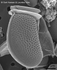
Description Large bag-shaped cell with well-developed sulcal lists. Cells 60–80 μm in length and 33–40 μm in dorso-ventral depth. Cells long and subovate ending in a broadly rounded posterior (a dorsal bulge). The posterior end is the widest. Left sulcal list well developed and very long; it can extend up to 4/5 of the cell. Thick thecal plates of the hypotheca deeply areolated, each areola with a pore. Small form of D. fortii considered for a long time as a distinct species (D. lapidistrigiliformis). [details] Description Medium-sized cell, broadly subovoid, widest posteriorly. Dorsal margin curved and ventral margin almost straight. Left...
Description Medium-sized cell, broadly subovoid, widest posteriorly. Dorsal margin curved and ventral margin almost straight. Left sulcal list long and can be up to four-fifths of the cell length. Right sulcal list also well developed and can extend beyond the Rl. Surface with deep poroids, each with a pore. Surface markings of type E. [details] Distribution neritic and oceanic; cold temperate to tropical waters worldwide
Distribution neritic and oceanic; cold temperate to tropical waters worldwide [details]
Guiry, M.D. & Guiry, G.M. (2024). AlgaeBase. World-wide electronic publication, National University of Ireland, Galway (taxonomic information republished from AlgaeBase with permission of M.D. Guiry). Dinophysis fortii Pavillard, 1924. Accessed through: World Register of Marine Species at: https://www.marinespecies.org/aphia.php?p=taxdetails&id=109624 on 2024-07-26
Date action by 2006-07-20 06:41:05Z changed Camba Reu, Cibran Copyright notice: the information originating from AlgaeBase may not be downloaded or replicated by any means, without the written permission of the copyright owner (generally AlgaeBase). Fair usage of data in scientific publications is permitted.
original description
Pavillard J. 1916. Recherches sur les Péridiniens du Golfe du Lion. <i>Trav. Inst. Bot. Univ. Montpellier </i>4: 9-70. [details]
original description (of Dinophysis lapidistrigiliformis Abé, 1967) Abé, T.H. (1967). The armoured Dinoflagellata: II. Prorocentridae and Dinophysidae (B) - <i>Dinophysis</i> and its allied genera. <em>Publications of the Seto Marine Biological Laboratory.</em> 2: 37-78. [details] Available for editors basis of record Guiry, M.D. & Guiry, G.M. (2024). AlgaeBase. <em>World-wide electronic publication, National University of Ireland, Galway.</em> searched on YYYY-MM-DD., available online at http://www.algaebase.org [details] basis of record Gómez, F. (2005). A list of free-living dinoflagellate species in the world's oceans. <em>Acta Bot. Croat.</em> 64(1): 129-212. [details] additional source Guiry, M.D. & Guiry, G.M. (2024). AlgaeBase. <em>World-wide electronic publication, National University of Ireland, Galway.</em> searched on YYYY-MM-DD., available online at http://www.algaebase.org [details] additional source Integrated Taxonomic Information System (ITIS). , available online at http://www.itis.gov [details] additional source Tomas, C.R. (Ed.). (1997). Identifying marine phytoplankton. Academic Press: San Diego, CA [etc.] (USA). ISBN 0-12-693018-X. XV, 858 pp., available online at http://www.sciencedirect.com/science/book/9780126930184 [details] additional source Brandt, S. (2001). Dinoflagellates, <B><I>in</I></B>: Costello, M.J. <i>et al.</i> (Ed.) (2001). <i>European register of marine species: a check-list of the marine species in Europe and a bibliography of guides to their identification. Collection Patrimoines Naturels,</i> 50: pp. 47-53 (look up in IMIS) [details] additional source Horner, R. A. (2002). A taxonomic guide to some common marine phytoplankton. <em>Biopress Ltd. Bristol.</em> 1-195. [details] additional source Abé, T.H. (1967). The armoured Dinoflagellata: II. Prorocentridae and Dinophysidae (B) - <i>Dinophysis</i> and its allied genera. <em>Publications of the Seto Marine Biological Laboratory.</em> 2: 37-78. [details] Available for editors additional source Yasumoto T., Oshima Y., Sugawara W., Fukuyo Y., Oguri H., Igarashi T. & Fujita N. 1980. Identification of <i>Dinophysis fortii</i> as the causative organism of diarrhetic shellfish poisoning. Bull. Jap. Soc. Sci. Fish. 46: 1405-1411. [details] additional source Lee J.S., lgarashi T., Fraga S., Dahl E., Hovgaard P. & Yasumoto T. (1989). Determination of diarrhetic shellfish toxins in various dinoflagellate species. <em>J. Appl. Phycol.</em> 1, 147-152. [details] additional source Suzuki T., Mitsuya T., Imai M. & Yamasaki M. 1997. DSP toxin contents in <i>Dinophysis fortii</i> and scallops collected at Mutsu Bay, Japan. J. Appl. Phycol. 8: 509-515.<br><br> Sato S., Koike K. & Kodama M. 1996. Seasonal variation of okadaic acid and dinophysistoxin-1 in <i>Dinophysis</i> spp. in association with the toxicity of scallop. In: <i>Harmful and Toxic Algal Blooms</i> (Ed. by T. Yasumoto, Y. Oshima & Y. Fukuyo), pp. 285-288. IOC, UNESCO, Paris. [details] additional source Steidinger, K. A., M. A. Faust, and D. U. Hernández-Becerril. 2009. Dinoflagellates (Dinoflagellata) of the Gulf of Mexico, Pp. 131–154 in Felder, D.L. and D.K. Camp (eds.), Gulf of Mexico–Origins, Waters, and Biota. Biodiversity. Texas A&M Press, College [details] additional source Balech, E. (1962). Tintinnoinea y Dinoflagellata del Pacífico según material de las expediciones Norpac y Downwind del Instituto Scripps de Oceanografía. <em>Rev. Mus. Arg. Cs. Nat. “B. Rivadavia”, C. Zool.</em> 7(1): 1-253, 26 pl. [details] Available for editors additional source Liu, J.Y. [Ruiyu] (ed.). (2008). Checklist of marine biota of China seas. <em>China Science Press.</em> 1267 pp. (look up in IMIS) [details] Available for editors additional source Lakkis, S. (2011). Le phytoplancton marin du Liban (Méditerranée orientale): biologie, biodiversité, biogéographie. Aracne: Roma. ISBN 978-88-548-4243-4. 293 pp. (look up in IMIS) [details] additional source Chang, F.H.; Charleston, W.A.G.; McKenna, P.B.; Clowes, C.D.; Wilson, G.J.; Broady, P.A. (2012). Phylum Myzozoa: dinoflagellates, perkinsids, ellobiopsids, sporozoans, in: Gordon, D.P. (Ed.) (2012). New Zealand inventory of biodiversity: 3. Kingdoms Bacteria, Protozoa, Chromista, Plantae, Fungi. pp. 175-216. [details] additional source Steidinger, K.A.; Tangen, K. (1997). Dinoflagellates. pp. 387-584. In: C.R. Tomas (ed.) (1997). Identifying Marine Phytoplankton. Academic Press: San Diego, CA [etc.] (USA). ISBN 0-12-693018-X. XV, 858 pp., available online at http://www.sciencedirect.com/science/article/pii/B9780126930184500057 [details] additional source Kofoid, C.A.; Skogsberg, T. (1928). Reports on the scientific results of the expedition to the Eastern Tropical Pacific, in charge of Alexander Agassiz, by the U.S. Fish Commission Steamer "Albatross" from October 1904 to March 1905, Lieut. Commander L.M. Garrett, U.S.N., Commanding. [No.] XXXV. The Dinoflagellata: the Dinophysoidae. <em>Memoirs of the Museum of Comparative Zoölogy, at Harvard College, Cambridge, Mass.</em> 51: 1-766., available online at https://www.biodiversitylibrary.org/page/4365822 [details] Available for editors additional source Balech, E. (2002). Dinoflagelados tecados tóxicos en el Cono Sur Americano. <em>In: Sar, E.A., Ferrario, M.E. & Reguera, B. (Eds.). Floraciones Algales Nocivas en el Cono Sur Americano. Instituto Español de Oceanografía.</em> pp. 123-144. [details] Available for editors new combination reference Pavillard J. 1923. A propos de la systématique des Péridiniens. Bull. Soc. Bot. France 70: 876-882. [details] ecology source Ishimaru, T.; Inoue, H.; Fukuyo, Y.; Ogata, T.; Kodama, M. (1988). Cultures of Dinophysis fortii and D. acuminata with the cryptomonad, Plagioselmis sp. <em>Mycotoxins.</em> 1988(1Supplement): 19-20., available online at https://doi.org/10.2520/myco1975.1988.1supplement_19 [details] ecology source Leles, S. G.; Mitra, A.; Flynn, K. J.; Stoecker, D. K.; Hansen, P. J.; Calbet, A.; McManus, G. B.; Sanders, R. W.; Caron, D. A.; Not, F.; Hallegraeff, G. M.; Pitta, P.; Raven, J. A.; Johnson, M. D.; Glibert, P. M.; Våge, S. (2017). Oceanic protists with different forms of acquired phototrophy display contrasting biogeographies and abundance. <em>Proceedings of the Royal Society B: Biological Sciences.</em> 284(1860): 20170664., available online at https://doi.org/10.1098/rspb.2017.0664 [details] Available for editors ecology source Mitra, A.; Caron, D. A.; Faure, E.; Flynn, K. J.; Leles, S. G.; Hansen, P. J.; McManus, G. B.; Not, F.; Do Rosario Gomes, H.; Santoferrara, L. F.; Stoecker, D. K.; Tillmann, U. (2023). The Mixoplankton Database (MDB): Diversity of photo‐phago‐trophic plankton in form, function, and distribution across the global ocean. <em>Journal of Eukaryotic Microbiology.</em> 70(4)., available online at https://doi.org/10.1111/jeu.12972 [details] ecology source Nagai, S.; Nitshitani, G.; Tomaru, Y.; Sakiyama, S.; Kamiyama, T. (2008). Predation by the toxic dinoflagellate <i>Dinophysis fortii</i> on the ciliate <i>Myrionecta rubra</i> and observation of sequestration of ciliate chloroplasts1. <em>Journal of Phycology.</em> 44(4): 909-922., available online at https://doi.org/10.1111/j.1529-8817.2008.00544.x [details]  Present Present  Present in aphia/obis/gbif/idigbio Present in aphia/obis/gbif/idigbio  Inaccurate Inaccurate  Introduced: alien Introduced: alien  Containing type locality Containing type locality
From editor or global species database
LSID urn:lsid:algaebase.org:taxname:47046 [details]From regional or thematic species database
Description Large bag-shaped cell with well-developed sulcal lists. Cells 60–80 μm in length and 33–40 μm in dorso-ventral depth. Cells long and subovate ending in a broadly rounded posterior (a dorsal bulge). The posterior end is the widest. Left sulcal list well developed and very long; it can extend up to 4/5 of the cell. Thick thecal plates of the hypotheca deeply areolated, each areola with a pore. Small form of D. fortii considered for a long time as a distinct species (D. lapidistrigiliformis).[details] Description Medium-sized cell, broadly subovoid, widest posteriorly. Dorsal margin curved and ventral margin almost straight. Left sulcal list long and can be up to four-fifths of the cell length. Right sulcal list also well developed and can extend beyond the Rl. Surface with deep poroids, each with a pore. Surface markings of type E. [details] Distribution neritic and oceanic; cold temperate to tropical waters worldwide [details] Harmful effect The first species of Dinophysis identified as the causative agent of DSP outbreaks(Yasumoto et al. 1980). Producer of okadaic acid (OA), dinophysis toxins (DTX1)and pectenotoxins (PTX2), toxins implicated in DSP. The main agent of DSP outbreaks in Japan and the Adriatic Sea. Accompanies other species of Dinophysis during DSP events in many parts of the world. Analyses of picked cells by HPLC-FD showed some Japanese strains contained OA (23 pg/cell) and other contained very high levels of DTX1 (13-191.5 pg/cell) and PTX2 (42.5 pg/cell) (Lee et al. 1989). More recent analyses by LC-MS showed cells containing DTX1 (8-11 pg/cell) and PTX2 (51-64 pg/cell) (Suzuki et al. 2009). Populations from the Adriatic Sea had dominance of PTX but also contained OA (15 pg/cell) (Draisci et al. 1996). [details] Introduced species impact Chinese part of the Yellow Sea (Marine Region) Other impact - undefined or uncertain (Bloom forming) [details] Introduced species vector dispersal Chinese part of the Yellow Sea (Marine Region) Ships: General [details] Synonymy Apparently no recent synonym other than Dinophysis lapidistrigiliformis Abé, reported by some authors as 'small cells' in the life cycle of D. fortii (Fukuyo et al. 1981, Otsuchi Mar. Res. Cent., Rep. 7: 3-23; Uchida et al. 1999, Bull. Fish. Environ. Inland Sea 1: 163-165). [details] From other sources
Diet general for group: both heterotrophic (eat other organisms) and autotrophic (photosynthetic) [details]Habitat pelagic [details] Importance General: known for producing dangerous toxins, particularly when in large numbers, called "red tides" because the cells are so abundant they make water change color. Also they can produce non-fatal or fatal amounts of toxins in predators (particularly shellfish) that may be eaten by humans. [details] Predators marine microorganisms and animal larvae [details] Reproduction general for group: both sexual and asexual [details]
Published in AlgaeBase
 Published in AlgaeBase  (from synonym Dinophysis lapidistrigiliformis Abé, 1967) (from synonym Dinophysis lapidistrigiliformis Abé, 1967)Published in AlgaeBase  (from synonym Dinophysis ovum F.Schütt, 1895) (from synonym Dinophysis ovum F.Schütt, 1895)Published in AlgaeBase  (from synonym Dinophysis brevisulca Tai & Skogsberg, 1934) (from synonym Dinophysis brevisulca Tai & Skogsberg, 1934)Published in AlgaeBase  (from synonym Dinophysis intermedia Pavillard, 1916) (from synonym Dinophysis intermedia Pavillard, 1916)To Barcode of Life To Biodiversity Heritage Library (8 publications) To Dyntaxa (from synonym Dinophysis ovum Schütt, 1895 sensu Martin, 1929) To European Nucleotide Archive, ENA (Dinophysis brevisulcus) (from synonym Dinophysis brevisulcus Tai & Skogsberg, 1934) To European Nucleotide Archive, ENA (Dinophysis fortii) To European Nucleotide Archive, ENA (Dinophysis ovum) (from synonym Dinophysis ovum F.Schütt, 1895) To European Nucleotide Archive, ENA (Dinophysis ovum) (from synonym Dinophysis ovum Schütt, 1895 sensu Martin, 1929) To GenBank (1 nucleotides; 0 proteins) (from synonym Dinophysis brevisulcus Tai & Skogsberg, 1934) To GenBank (88 nucleotides; 1 proteins) To PESI To PESI (from synonym Dinophysis ovum Schütt, 1895 sensu Martin, 1929) To PESI (from synonym Dinophysis ovum F.Schütt, 1895) To ITIS |

