Photogallery

G. guangdongensis
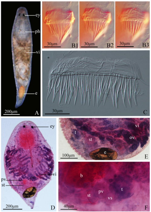
Description A) Photographed in life. (B) Sclerotic stylet in whole-mounted specimen (1, 2, 3 represent different depth of microscope view). (C) Dissection of sclerotic stylet. (D) Permanent slide stained by H.E. method. (E) Detail of reproductive system stained by H. E. method. (F)Ventral view of the male copulatory organ stained by H.E. method.
PNG file - 1.07 MB - 800 x 1 124 pixels
added on 2017-04-07842 viewsWoRMS taxaScan of photo Gieysztoria guangdongensis Wang & Xia, 2013checked Tyler, Seth 2017-04-07
This work is licensed under a Creative Commons Attribution-NonCommercial-ShareAlike 4.0 International License
Click here to return to the thumbnails overview
