
Photogallery

P. gilberti
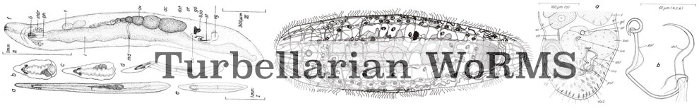
Description FIGURE 4. Phaenocora gilberti sp. nov. A, habitus of live animal. B1, horizontal reconstruction of the genital system; B2, section through the burso-intestinal duct, which connects to the intestinal bursa from the dorsal side at the place indicated?? in figure B1; B3 reconstruction of the superior genital atrium, from which both the male and female system dorsally departs at the place indicated?? in figure B1. The structure indicated with gl4 are the cell bodies of the glands gl4, which enter the female genital canal more dorsally (not indicated in the figures); C, reproductive system of live animal. D, schematic representation of the cirrus. (B, D: SMNH Type-8672).
PNG file - 426.63 kB - 800 x 1 000 pixels
added on 2017-04-07736 viewsWoRMS taxaScan of photo Phaenocora gilberti Houben & Artois, 2014checked Tyler, Seth 2017-04-07
This work is licensed under a Creative Commons Attribution-NonCommercial-ShareAlike 4.0 International License
Click here to return to the thumbnails overview