Scleractinia name details
Paraplacocoenia Beauvais, 1982 †
1463432 (urn:lsid:marinespecies.org:taxname:1463432)
unaccepted (synonymy)
Genus
Placocoenia orbignyana Reuss, 1854 † accepted as Placocoeniopsis orbignyana (Reuss, 1854) † (type by original designation)
- Species Paraplacocoenia orbignyana (Reuss, 1854) † accepted as Placocoeniopsis orbignyana (Reuss, 1854) † (changed combination)
marine, fresh, terrestrial
fossil only
Beauvais M. (1982). Révision Systématique des Madréporaires des couches de Gosau (Crétacé supérieur, Autriche). 5 vols. <em>Paris (Comptoir géologique). - Trav. Lab. Paleont. Univ. P. & M. Curie. 1-710. Paris.</em> [details]
Hoeksema, B. W.; Cairns, S. (2021). World List of Scleractinia. Paraplacocoenia Beauvais, 1982 †. Accessed at: https://marinespecies.org/scleractinia/aphia.php?p=taxdetails&id=1463432 on 2025-04-27
original description
Beauvais M. (1982). Révision Systématique des Madréporaires des couches de Gosau (Crétacé supérieur, Autriche). 5 vols. <em>Paris (Comptoir géologique). - Trav. Lab. Paleont. Univ. P. & M. Curie. 1-710. Paris.</em> [details]
From editor or global species database
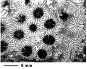
Diagnosis Colonial, massive or knobby, plocoid. Gemmation due to extracalicinal budding. Corallites united by a granulate peritheca. Costosepta radial, granular laterally, compact, in the costal area dissociating into trabecular structures. Septal margins beaded. Peritheca tabulo-columnar. Small trabecular–lamellar columella. Endothecal dissepiments thin and subtabulate, abundant. Wall septoparathecal. Septal microstructure consisting of simple trabeculae forming dark axial lines. [details]Remark In many respects the genus Paraplacocoenia Beauvais resembles the genus Haldonia. However, very granulate peritheca, dissociating into trabecular structures in the costal area and a lamellar columella are present in Paraplacocoenia but not in Haldonia. Furthermore, based on closely corresponding features, the genus Paraplacocoenia was merged with the Neocoenia as the subgenus of the latter (Baron-Szabo, 2014). Also see information under Hyndophorposis Soehle, 1899. [details]
