
WoRMS Photogallery
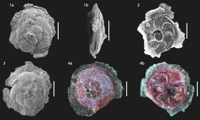
Rotaliammina chitinosa (Collins, 1958), specimens from Brazil. Scale bar = 100 μm. SOURCE: Disaró et al. 2022.
Description 1a and 1b) SEM views of the same specimen - spiral side and profile, respectively. 2) SEM umbilical view; note the collapsed chambers and the axial depression. 3) SEM spiral view; note the peripheral flange, by means of which the test adheres to the substrate. 4a and 4b) Views of the same specimen under light microscopy- spiral side and umbilical side, respectively; note the flange at the peripheral margin, which maintains adhesion to the substrate, and the collapsed chambers on the umbilical side.
PNG file - 380.31 kB - 710 x 427 pixels
added on 2026-09-2917 viewsWoRMS taxaScan of photo Rotaliammina chitinosa (Collins, 1958)checked Trevisan Disaró, Sibelle 2026-09-29Source:
Disaró, S. T.; Totah, V. I.; Watanabe, S.; Ribas, E. R.; Pupo, D. V. (2022). Biodiversidade ...

This work is licensed under a Creative Commons Attribution-NonCommercial-ShareAlike 4.0 International License
Click here to return to the thumbnails overview
Comment (0)
Click here to add a comment.* indicates a required field.
Disclaimer: WoRMS does not exercise any editorial control over the information displayed here. However, if you come across any misidentifications, spelling mistakes or low quality pictures, your comments would be very much appreciated. You can reach us by emailing info@marinespecies.org or adding a comment, we will correct the information or remove the image from the website when necessary or in case of doubt.
