Photogallery

Tripaphylus squidwardi Boxshall, Barton, Kirke, Zhu & Johnson, 2022
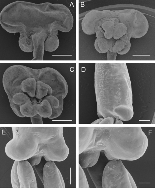
Description Fig. 2 Scanning electron micrographs of Tripaphylus squidwardi n. sp. adult female. A, cephalothorax, dorsal; B, cephalothorax, ventral; C, cephalothorax of another specimen, ventral; D, Posterior extremity of trunk, lateral view showing lateral lobe at posterior end of trunk; E, Posterior extremity of trunk, showing origins of posterior processes, egg sacs and abdomen; F, abdomen, showing anal prominence. Scale bars: A-C, 1.0 mm, D-E, 500 lm, F, 200 lm
Source: Boxshall, G. A.; Barton, D. P.; Kirke, A.; Zhu, X.; Johnson, G. (2022). Two new parasitic copepods of the family Sphyriidae (Copepoda: Siphonostomatoida) from Australian elasmobranchs. Systematic Parasitology. 99(6): 659-669., available online at https://doi.org/10.1007/s11230-022-10054-4
·
This work is licensed under a Creative Commons Attribution-NonCommercial-ShareAlike 4.0 International License
Click here to return to the thumbnails overview

