WoRMS Photogallery
P. lutheri
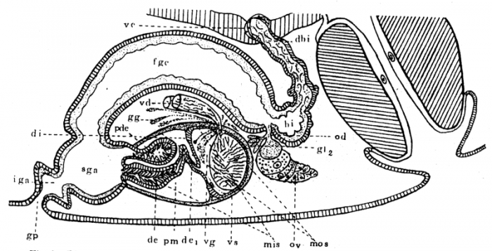
Description Diagrammatic reconstruction of the genital organs of Phaenocora lutheri n. sp. Note pseudo-ductus ejaculatorius (pde), and the division of the muscles of the male copulatory organ into those derived from the primitive inner sac (mis) and those derived from outer sac (mos).
PNG file - 122.36 kB - 800 x 409 pixels
added on 2017-04-07446 viewsWoRMS taxaScan of photo Phaenocora lutheri Gilbert, 1937checked Tyler, Seth 2017-04-07
Click here to return to the thumbnails overview
 Comment (0)
 Click here to add a comment.
* indicates a required field.
Disclaimer: WoRMS does not exercise any editorial control over the information displayed here. However, if you come across any misidentifications, spelling mistakes or low quality pictures, your comments would be very much appreciated. You can reach us by emailing info@marinespecies.org or adding a comment, we will correct the information or remove the image from the website when necessary or in case of doubt.
