WoRMS taxon details
Convoluta borealis Sabussow, 1900
142600 (urn:lsid:marinespecies.org:taxname:142600)
accepted
Species
Childia groenlandica borealis Steinböck, 1932 · unaccepted (homonym & junior synonym)
marine
Sabussow, H. (1900). Beobachtungen über die Turbellarien der Inseln von Salowetzk. <em>Trudy Obshchestva yestestvoispytateley pri Imperatorskom Kazanskom universitete (= Proceedings of the Society of Naturalists at the Imperial Kazan University).</em> 34(4): 1–208. 3 plates.
page(s): 3 [details]
page(s): 3 [details]
Type locality contained in Russia
type locality contained in Russia [details]
Description "....Adjacent to the ovary, on its inner side, there is a cluster of dark-pigmented cells on each side, likely glandular in...
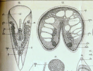
Description “5. *Convoluta borealis* n. sp. (Pl. II, figs. 14, 15).
Size 1–4 mm; the body is broad and flat, with a rounded...
Description "....Adjacent to the ovary, on its inner side, there is a cluster of dark-pigmented cells on each side, likely glandular in nature. The *bursa seminalis* consists of a thin-walled sac with a fibrous structure—filled with spermatozoa—and a chitinous tip. The chitinous tip appears to be composed of separate, relatively broad chitinous rings stacked upon one another; these exhibit radial striations and are perforated by a canal whose diameter decreases with increasing distance from the *bursa seminalis*. A thick sheath surrounds the chitinous tip, with nuclei situated at its periphery; this sheath most likely originates from the cells of the *matrix* involved in the formation of the chitinous tube. The outer opening of the chitinous tube faces anteriorly. Among the male reproductive organs, I was able to observe the penis, which lacks chitin and lies at the posterior end of the body, not far from the genital opening. The penis is shaped like a spherical sac with muscular walls and is enclosed in a special pocket (the *Penistasche* or *Penisscheide* of German authors) with fibrous walls.
The female genital opening is situated between the *bursa seminalis* and the penis—specifically, in the opening below the penis.
Distribution. Found throughout the area around the Solovetsky Islands among algae at shallow depths; sometimes under stones near the shore. 1895, 1897.”
[details]
The female genital opening is situated between the *bursa seminalis* and the penis—specifically, in the opening below the penis.
Distribution. Found throughout the area around the Solovetsky Islands among algae at shallow depths; sometimes under stones near the shore. 1895, 1897.”
[details]
Description “5. *Convoluta borealis* n. sp. (Pl. II, figs. 14, 15).
Size 1–4 mm; the body is broad and flat, with a rounded...
Description “5. *Convoluta borealis* n. sp. (Pl. II, figs. 14, 15).
Size 1–4 mm; the body is broad and flat, with a rounded anterior end, gradually tapering toward the posterior end, which forms a sharp, tail-like projection. The lateral margins curve toward the ventral side, forming prominent folds. The body color is brownish-green or olive-yellow, resulting from the presence of unicellular yellow and green algae in the parenchyma (yellow-green algae are the most frequently encountered). The algae are spherical or oval and contain a vacuole.
The epithelium consists of flat, plate-like, polygonal cells—with a height of 0.003 mm—containing elongated-oval nuclei. The nuclei are spaced widely apart due to the relatively great width of the epithelial cellular elements and are oriented such that their long axis lies parallel to the outer surface of the epithelium, while their short axis is perpendicular to it.
Under high magnification, the cytoplasm of the epithelial cells appears striated parallel to the cell height. The epithelial surface is bounded by a cuticle that shares the same structure as that of other Acoela—namely, consisting of the bases of the cilia (*Fussstücke der Cilien*). The cilia are colorless and measure 0.004 mm in length, making them nearly twice as tall as the epithelium itself.
The body wall consists of four muscle layers, none of which, however, is particularly strongly developed. These muscle layers are arranged as follows: outermost lies a layer of circular fibers, followed by two diagonal layers, while the innermost layer consists of longitudinal fibers—the most developed of the group—arranged in several rows.
Numerous pear-shaped glands open onto the epithelium; the bodies of these glands are situated in the parenchyma, well below the layers of the body wall.
The parenchyma is structured similarly to that of *Convoluta sordida* (according to von Graff, 20): it resembles reticular tissue in the anterior, posterior, and lateral regions, whereas the central part of the body is filled with a plasmodium-like tissue responsible for digesting food. Dorsoventral muscle fibers are particularly well-developed in the anterior, posterior, and lateral regions, while being sparse or absent in the center of the body. Two poison organs, each consisting of a spherical body and a cylindrical chitinous tube, are located in the parenchyma anterior to the ovaries.
The nervous system is structured similarly to that of other representatives of the genus *Convoluta*. The brain is arc-shaped; six pairs of nerves emerge from it, interconnected by anastomoses. The structure of the otocyst is identical to that found in other representatives of the genus *Convoluta*. The reproductive organs of *Convoluta borealis* are differentiated into male and female systems.
The female reproductive organs consist of 1) paired ovaries and 2) a *bursa seminalis*, whereas the male reproductive organs consist of 2) testes and 2) a copulatory organ.
The ovaries begin at the start of the second third of the body; in this region, they consist of several rows of cells characterized by a large, vesicular nucleus with a nucleolus and granular, lustrous protoplasm. Towards the animal's posterior end, the ova are arranged in a single row and gradually increase in size. The largest ova—measuring 0.114 mm in diameter—are located at the end of the ovary, whereas the small ova in the anterior section measure only 0.022 mm in diameter; their nuclei measure 0.012 mm, and the nucleolus 0.004 mm. The large eggs—typically two in number—possess nuclei shaped like the figure 8 or a gymnastic weight (evidently ready to divide) and lie within distinct cavities. No sharply defined *tunica propria* can be observed around the ovary or the individual eggs....."
[details]
Size 1–4 mm; the body is broad and flat, with a rounded anterior end, gradually tapering toward the posterior end, which forms a sharp, tail-like projection. The lateral margins curve toward the ventral side, forming prominent folds. The body color is brownish-green or olive-yellow, resulting from the presence of unicellular yellow and green algae in the parenchyma (yellow-green algae are the most frequently encountered). The algae are spherical or oval and contain a vacuole.
The epithelium consists of flat, plate-like, polygonal cells—with a height of 0.003 mm—containing elongated-oval nuclei. The nuclei are spaced widely apart due to the relatively great width of the epithelial cellular elements and are oriented such that their long axis lies parallel to the outer surface of the epithelium, while their short axis is perpendicular to it.
Under high magnification, the cytoplasm of the epithelial cells appears striated parallel to the cell height. The epithelial surface is bounded by a cuticle that shares the same structure as that of other Acoela—namely, consisting of the bases of the cilia (*Fussstücke der Cilien*). The cilia are colorless and measure 0.004 mm in length, making them nearly twice as tall as the epithelium itself.
The body wall consists of four muscle layers, none of which, however, is particularly strongly developed. These muscle layers are arranged as follows: outermost lies a layer of circular fibers, followed by two diagonal layers, while the innermost layer consists of longitudinal fibers—the most developed of the group—arranged in several rows.
Numerous pear-shaped glands open onto the epithelium; the bodies of these glands are situated in the parenchyma, well below the layers of the body wall.
The parenchyma is structured similarly to that of *Convoluta sordida* (according to von Graff, 20): it resembles reticular tissue in the anterior, posterior, and lateral regions, whereas the central part of the body is filled with a plasmodium-like tissue responsible for digesting food. Dorsoventral muscle fibers are particularly well-developed in the anterior, posterior, and lateral regions, while being sparse or absent in the center of the body. Two poison organs, each consisting of a spherical body and a cylindrical chitinous tube, are located in the parenchyma anterior to the ovaries.
The nervous system is structured similarly to that of other representatives of the genus *Convoluta*. The brain is arc-shaped; six pairs of nerves emerge from it, interconnected by anastomoses. The structure of the otocyst is identical to that found in other representatives of the genus *Convoluta*. The reproductive organs of *Convoluta borealis* are differentiated into male and female systems.
The female reproductive organs consist of 1) paired ovaries and 2) a *bursa seminalis*, whereas the male reproductive organs consist of 2) testes and 2) a copulatory organ.
The ovaries begin at the start of the second third of the body; in this region, they consist of several rows of cells characterized by a large, vesicular nucleus with a nucleolus and granular, lustrous protoplasm. Towards the animal's posterior end, the ova are arranged in a single row and gradually increase in size. The largest ova—measuring 0.114 mm in diameter—are located at the end of the ovary, whereas the small ova in the anterior section measure only 0.022 mm in diameter; their nuclei measure 0.012 mm, and the nucleolus 0.004 mm. The large eggs—typically two in number—possess nuclei shaped like the figure 8 or a gymnastic weight (evidently ready to divide) and lie within distinct cavities. No sharply defined *tunica propria* can be observed around the ovary or the individual eggs....."
[details]
Tyler, S., Artois, T.; Schilling, S.; Hooge, M.; Bush, L.F. (eds) (2006-2026). World List of turbellarian worms: Acoelomorpha, Catenulida, Rhabditophora. Convoluta borealis Sabussow, 1900. Accessed through: World Register of Marine Species at: https://www.marinespecies.org/aphia.php?p=taxdetails&id=142600 on 2026-09-13
Date
action
by
 The webpage text is licensed under a Creative Commons
Attribution 4.0 License
The webpage text is licensed under a Creative Commons
Attribution 4.0 License
Nomenclature
original description
Sabussow, H. (1900). Beobachtungen über die Turbellarien der Inseln von Salowetzk. <em>Trudy Obshchestva yestestvoispytateley pri Imperatorskom Kazanskom universitete (= Proceedings of the Society of Naturalists at the Imperial Kazan University).</em> 34(4): 1–208. 3 plates.
page(s): 3 [details]
basis of record Faubel, A.; Noreña, C. (2001). Turbellaria, <B><I>in</I></B>: Costello, M.J. <i>et al.</i> (Ed.) (2001). <i>European register of marine species: a check-list of the marine species in Europe and a bibliography of guides to their identification. Collection Patrimoines Naturels,</i> 50: pp. 123-136 (look up in IMIS) [details] Available for editors [request]
page(s): 3 [details]
basis of record Faubel, A.; Noreña, C. (2001). Turbellaria, <B><I>in</I></B>: Costello, M.J. <i>et al.</i> (Ed.) (2001). <i>European register of marine species: a check-list of the marine species in Europe and a bibliography of guides to their identification. Collection Patrimoines Naturels,</i> 50: pp. 123-136 (look up in IMIS) [details] Available for editors
[request]Other
additional source
van der Land, J. (ed). (2008). UNESCO-IOC Register of Marine Organisms (URMO). , available online at http://www.marinespecies.org/urmo/ [details]
 Present Present in aphia/obis/gbif/idigbio Inaccurate Introduced: alien Containing type locality
From editor or global species database
Description "....Adjacent to the ovary, on its inner side, there is a cluster of dark-pigmented cells on each side, likely glandular in nature. The *bursa seminalis* consists of a thin-walled sac with a fibrous structure—filled with spermatozoa—and a chitinous tip. The chitinous tip appears to be composed of separate, relatively broad chitinous rings stacked upon one another; these exhibit radial striations and are perforated by a canal whose diameter decreases with increasing distance from the *bursa seminalis*. A thick sheath surrounds the chitinous tip, with nuclei situated at its periphery; this sheath most likely originates from the cells of the *matrix* involved in the formation of the chitinous tube. The outer opening of the chitinous tube faces anteriorly. Among the male reproductive organs, I was able to observe the penis, which lacks chitin and lies at the posterior end of the body, not far from the genital opening. The penis is shaped like a spherical sac with muscular walls and is enclosed in a special pocket (the *Penistasche* or *Penisscheide* of German authors) with fibrous walls. The female genital opening is situated between the *bursa seminalis* and the penis—specifically, in the opening below the penis.
Distribution. Found throughout the area around the Solovetsky Islands among algae at shallow depths; sometimes under stones near the shore. 1895, 1897.”
[details]
Description “5. *Convoluta borealis* n. sp. (Pl. II, figs. 14, 15).
Size 1–4 mm; the body is broad and flat, with a rounded anterior end, gradually tapering toward the posterior end, which forms a sharp, tail-like projection. The lateral margins curve toward the ventral side, forming prominent folds. The body color is brownish-green or olive-yellow, resulting from the presence of unicellular yellow and green algae in the parenchyma (yellow-green algae are the most frequently encountered). The algae are spherical or oval and contain a vacuole.
The epithelium consists of flat, plate-like, polygonal cells—with a height of 0.003 mm—containing elongated-oval nuclei. The nuclei are spaced widely apart due to the relatively great width of the epithelial cellular elements and are oriented such that their long axis lies parallel to the outer surface of the epithelium, while their short axis is perpendicular to it.
Under high magnification, the cytoplasm of the epithelial cells appears striated parallel to the cell height. The epithelial surface is bounded by a cuticle that shares the same structure as that of other Acoela—namely, consisting of the bases of the cilia (*Fussstücke der Cilien*). The cilia are colorless and measure 0.004 mm in length, making them nearly twice as tall as the epithelium itself.
The body wall consists of four muscle layers, none of which, however, is particularly strongly developed. These muscle layers are arranged as follows: outermost lies a layer of circular fibers, followed by two diagonal layers, while the innermost layer consists of longitudinal fibers—the most developed of the group—arranged in several rows.
Numerous pear-shaped glands open onto the epithelium; the bodies of these glands are situated in the parenchyma, well below the layers of the body wall.
The parenchyma is structured similarly to that of *Convoluta sordida* (according to von Graff, 20): it resembles reticular tissue in the anterior, posterior, and lateral regions, whereas the central part of the body is filled with a plasmodium-like tissue responsible for digesting food. Dorsoventral muscle fibers are particularly well-developed in the anterior, posterior, and lateral regions, while being sparse or absent in the center of the body. Two poison organs, each consisting of a spherical body and a cylindrical chitinous tube, are located in the parenchyma anterior to the ovaries.
The nervous system is structured similarly to that of other representatives of the genus *Convoluta*. The brain is arc-shaped; six pairs of nerves emerge from it, interconnected by anastomoses. The structure of the otocyst is identical to that found in other representatives of the genus *Convoluta*. The reproductive organs of *Convoluta borealis* are differentiated into male and female systems.
The female reproductive organs consist of 1) paired ovaries and 2) a *bursa seminalis*, whereas the male reproductive organs consist of 2) testes and 2) a copulatory organ.
The ovaries begin at the start of the second third of the body; in this region, they consist of several rows of cells characterized by a large, vesicular nucleus with a nucleolus and granular, lustrous protoplasm. Towards the animal's posterior end, the ova are arranged in a single row and gradually increase in size. The largest ova—measuring 0.114 mm in diameter—are located at the end of the ovary, whereas the small ova in the anterior section measure only 0.022 mm in diameter; their nuclei measure 0.012 mm, and the nucleolus 0.004 mm. The large eggs—typically two in number—possess nuclei shaped like the figure 8 or a gymnastic weight (evidently ready to divide) and lie within distinct cavities. No sharply defined *tunica propria* can be observed around the ovary or the individual eggs....."
[details]

To Biodiversity Heritage Library (4 publications)
To PESI
To the Turbellarian Taxonomic Database
To the Turbellarian Taxonomic Database
(from synonym Childia groenlandica borealis Steinböck, 1932)
To PESI
To the Turbellarian Taxonomic Database
To the Turbellarian Taxonomic Database
(from synonym Childia groenlandica borealis Steinböck, 1932)From editor or global species database