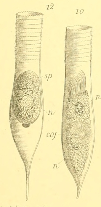
Amphorella fusiformis Meunier, 1919
Description Illustrations fro Meunier 1919, plate 22. Figure 10 show a specimen undergoing cell division. Figure 12 shows a specimen mostly likely in a cyst form or parasitized.
JPG file - 283.90 kB - 574 x 1 181 pixels
Extra information
FileName: 184902.jpg
FileDateTime: 1741600466
FileSize: 283899
FileType: 2
MimeType: image/jpeg
SectionsFound: ANY_TAG, IFD0, THUMBNAIL, EXIF
COMPUTED.html: width="574" height="1181"
COMPUTED.Height: 1181
COMPUTED.Width: 574
COMPUTED.IsColor: 1
COMPUTED.ByteOrderMotorola: 1
COMPUTED.Thumbnail.FileType: 2
COMPUTED.Thumbnail.MimeType: image/jpeg
Orientation: 1
XResolution: 3000000/10000
YResolution: 3000000/10000
ResolutionUnit: 2
Software: Adobe Photoshop CS5 Macintosh
DateTime: 2025:03:10 10:52:01
Exif_IFD_Pointer: 164
ColorSpace: 1
ExifImageWidth: 574
ExifImageLength: 1181
added on 2025-03-1055 viewsTraits taxa
FileName: 184902.jpg
FileDateTime: 1741600466
FileSize: 283899
FileType: 2
MimeType: image/jpeg
SectionsFound: ANY_TAG, IFD0, THUMBNAIL, EXIF
COMPUTED.html: width="574" height="1181"
COMPUTED.Height: 1181
COMPUTED.Width: 574
COMPUTED.IsColor: 1
COMPUTED.ByteOrderMotorola: 1
COMPUTED.Thumbnail.FileType: 2
COMPUTED.Thumbnail.MimeType: image/jpeg
Orientation: 1
XResolution: 3000000/10000
YResolution: 3000000/10000
ResolutionUnit: 2
Software: Adobe Photoshop CS5 Macintosh
DateTime: 2025:03:10 10:52:01
Exif_IFD_Pointer: 164
ColorSpace: 1
ExifImageWidth: 574
ExifImageLength: 1181
This work is licensed under a Creative Commons Attribution-NonCommercial-ShareAlike 4.0 International License
 Comment (0)