WoRMS image

Limnognathia maerski
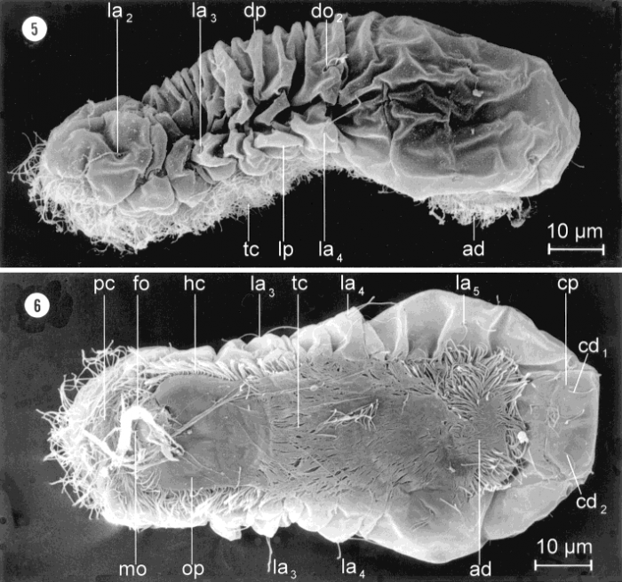
Description Limnognathia maerski nov. gen et nov. sp. Females. SEM. From culture, Copenhagen. Fig. 5: Lateral view of a slightly shrunken specimen showing the dorsal (dp) and lateral plates (lp). The locomotory cilia are ventral ciliation with trunk ciliophores (tc) and an adhesive ciliated pad (ad) posteriorly. Note the position of one of each of some of the paired sensoria: lateralia (la2-la4) and dorsalia (do2). Fig. 6: Ventral view of animal with anterior preoral cilia field (pc), head (hc) and trunk ciliophores (tc), and a posterior adhesive ciliated pad (ad). The mouth (mo) is partly covered by food (fo) or detritus and situated anterior to the oral plate (op). Note the position of some of the paired lateralia (la3-la5) and caudalia (cd1-cd2). cp, caudal plate.Source: https://doi.org/10.1002/1097-4687(200010)246:1<1::aid-jmor1>3.0.co;2-d PNG file - 149.76 kB - 711 x 666 pixels added on 2019-03-22180 viewsWoRMS taxaMicroscope Limnognathia maerski Kristensen & Funch, 2000checked Kristensen, Reinhardt Møbjerg 2019-03-22
This work is licensed under a Creative Commons Attribution-NonCommercial-ShareAlike 4.0 International License
 Comment (0)
Disclaimer: WoRMS does not exercise any editorial control over the information displayed here. However, if you come across any misidentifications, spelling mistakes or low quality pictures, your comments would be very much appreciated. You can reach us by emailing info@marinespecies.org, we will correct the information or remove the image from the website when necessary or in case of doubt.