Neoechiniscoides aski Møbjerg, Jørgensen & Kristensen, 2020
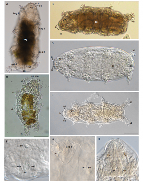
Description Figure 2. Light microscopy of Neoechiniscoides aski. A–C, images of live, moving specimens. A, ventral view of a live adult female, showing the anal system with lateral wings. In live animals, the conspicuous anal complex moves from side to side when the animal walks/runs. Scale bar: 50 µm. B, dorsal view of live adult female used for DNA extraction (28S GenBank accession number KX363645; COI GenBank accession number KX363656). Note the eight to nine mature oocytes. Scale bar: 50 µm. C, dorsal view of live adult male. Scale bar: 50 µm. D, holotype of N. aski (female). Scale bar: 50 µm. E, paratypic allotype of N. aski (male). Scale bar: 50 µm. F, ventrocaudal view of holotypic female showing anal system and the gonopore, which is raised on an elevation (note that seminal receptacles are lacking). Scale bar: 20 µm. G, ventral view of paratypicLink to publication: https://www.marinespecies.org/aphia.php?p=sourcedetails&id=391897
PNG file - 882.02 kB - 657 x 841 pixels added on 2021-03-15786 viewsURMO taxa
 Comment (0)