Asthenosoma (spines)
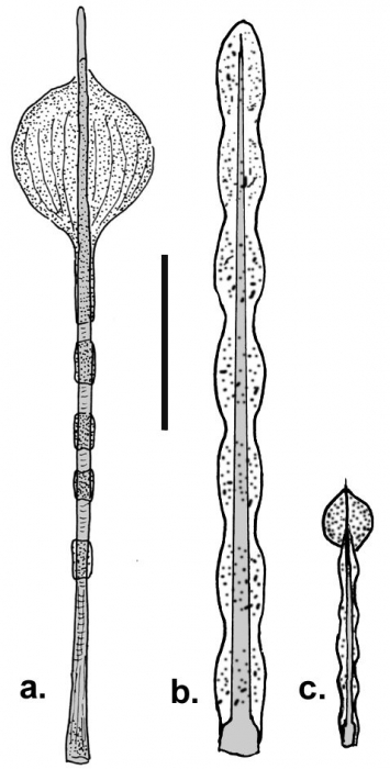
Description Venomous spines on the aboral side of Asthenosoma marisrubri and A. varium. a. Large secondary spine of A. marisrubri with a blister-like poison gland at the distal end, b. Large secondary spine of A. varium invested in a thick skin sheath annularly constricted and filled with venom. These are arranged in approximately rectangular areas (shaded in the ciagram below). This kind of spines is lacking in A. marisrubri, c. Small secondary spine of A. varium with a distal blister-like poison gland. These spines border the rectangular areas set with the large secondary spines (dots in the diagram below).Scale 3 mm. Modified from Weinberg & de Ridder (1998) JPG file - 57.27 kB - 478 x 945 pixels added on 2011-02-23281 viewsEchinoidea taxaScan of drawing Asthenosoma varium Grube, 1868checked Kroh, Andreas 2021-02-24Scan of drawing Asthenosoma marisrubri Weinberg & de Ridder, 1998checked Kroh, Andreas 2021-02-24From reference Schultz, H. (2011). Sea urchins III: Worldwide regular de... Download full size
 © 2011 Schultz, Heinke
 Comment (0)